颈椎病的临床诊断可能是由于患者的症状明显(例如颈部疼痛,颈椎神经根病或者脊髓型颈椎病)而发现,或者在评估其他症状时偶然发现。[7]Binder AI. Cervical spondylosis and neck pain. BMJ. 2007 Mar 10;334(7592):527-31.http://www.ncbi.nlm.nih.gov/pubmed/17347239?tool=bestpractice.com[16]Rao RD, Currier BL, Albert TJ, et al. Degenerative cervical spondylosis: clinical syndromes, pathogenesis and management. J Bone Joint Surg Am. 2007 Jun;89(6):1360-78.http://www.ncbi.nlm.nih.gov/pubmed/17575617?tool=bestpractice.com
对于颈部疼痛的患者,有一些症状应该视为红色警戒,一旦发现,应行进一步的检查,以排除重要的鉴别诊断。[16]Rao RD, Currier BL, Albert TJ, et al. Degenerative cervical spondylosis: clinical syndromes, pathogenesis and management. J Bone Joint Surg Am. 2007 Jun;89(6):1360-78.http://www.ncbi.nlm.nih.gov/pubmed/17575617?tool=bestpractice.com[26]Cook CE, Hegedus E, Pietrobon R, et al. A pragmatic neurological screen for patients with suspected cord compressive myelopathy. Phys Ther. 2007 Sep;87(9):1233-42.http://www.ncbi.nlm.nih.gov/pubmed/17636158?tool=bestpractice.com
近期高处坠落史或者头颈部外伤病史
无法解释的体重减轻
严重顽固性疼痛或者严重局部压痛
颈部淋巴结肿大
无法解释的发热,尤其是糖尿病患者
肿瘤病史
长期激素应用史。
主要表现为轴性颈椎痛的症状
轴性颈椎痛包括直接疼痛(尤其是颈后部疼痛)、牵涉痛或者间接痛。[2]Guzman J, Haldeman S, Carroll LJ, et al. Clinical practice implications of the Bone and Joint Decade 2000-2010 Task Force on Neck Pain and Its Associated Disorders: from concepts and findings to recommendations. J Manipulative Physiol Ther. 2009 Feb;32(2 suppl):S227-43.http://www.ncbi.nlm.nih.gov/pubmed/19251069?tool=bestpractice.com[6]Binder AI. Neck pain. BMJ Clin Evid. 2008 [internet publication].https://www.ncbi.nlm.nih.gov/pmc/articles/PMC2907992/http://www.ncbi.nlm.nih.gov/pubmed/19445809?tool=bestpractice.com[7]Binder AI. Cervical spondylosis and neck pain. BMJ. 2007 Mar 10;334(7592):527-31.http://www.ncbi.nlm.nih.gov/pubmed/17347239?tool=bestpractice.com 牵涉痛包括由于关节活动而引起椎旁肌肉痉挛、肌肉收缩及肌肉僵直引起的继发性疼痛。其他牵涉痛包括枕部疼痛和紧张性头痛。
轴性颈椎痛可存在于任一轴位颈部肌肉,包括斜角肌(前斜角肌综合征)、斜方肌、肩胛间肌及自枕骨至腰部区域的椎旁肌肉,这些都是轴位肌肉痉挛可以扩散到的区域。[20]Haldeman S, Carroll L, Cassidy JD. Findings from the bone and joint decade 2000 to 2010 task force on neck pain and its associated disorders. J Occup Environ Med. 2010 Apr;52(4):424-7.http://www.ncbi.nlm.nih.gov/pubmed/20357682?tool=bestpractice.com
颈椎病引起的颈部疼痛程度比体格检查得出的结果更具有主观性,体格检查可发现轻至中度椎旁肌肉痉挛、运动范围基本正常及椎旁肌群的疼痛(斜角肌、斜方肌、肩胛间肌及枕部肌肉等)。[2]Guzman J, Haldeman S, Carroll LJ, et al. Clinical practice implications of the Bone and Joint Decade 2000-2010 Task Force on Neck Pain and Its Associated Disorders: from concepts and findings to recommendations. J Manipulative Physiol Ther. 2009 Feb;32(2 suppl):S227-43.http://www.ncbi.nlm.nih.gov/pubmed/19251069?tool=bestpractice.com[6]Binder AI. Neck pain. BMJ Clin Evid. 2008 [internet publication].https://www.ncbi.nlm.nih.gov/pmc/articles/PMC2907992/http://www.ncbi.nlm.nih.gov/pubmed/19445809?tool=bestpractice.com[7]Binder AI. Cervical spondylosis and neck pain. BMJ. 2007 Mar 10;334(7592):527-31.http://www.ncbi.nlm.nih.gov/pubmed/17347239?tool=bestpractice.com
对于仅有单次发作的病例,治疗性试验可能足以明确诊断。没有必要进行进一步诊断性检查。
对于存在严重颈痛、慢性颈痛或者颈痛伴外伤或颈部手术史(近期或既往)的患者,应进行颈部 X 线检查。[5]American College of Radiology. ACR appropriateness criteria: cervical neck pain or cervical radiculopathy. 2018 [internet publication].https://acsearch.acr.org/docs/69426/Narrative/ 如果有恶性肿瘤或颈部手术史,指南指出,实施 MRI 更合适。[5]American College of Radiology. ACR appropriateness criteria: cervical neck pain or cervical radiculopathy. 2018 [internet publication].https://acsearch.acr.org/docs/69426/Narrative/
上肢放射性疼痛的症状表现(伴或不伴轴性颈椎痛)
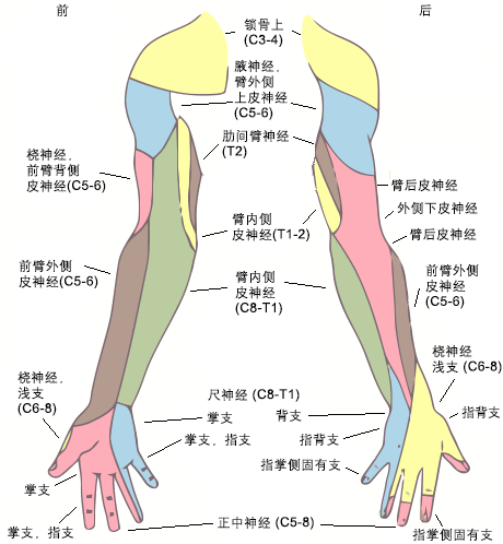
诊断神经根型颈椎病的依据:上肢放射性疼痛(肩部远端);累及 ≥1 个的皮节区域;特定神经根支配区的轻度肌力下降;以及单一水平的反射改变(如果存在)。[7]Binder AI. Cervical spondylosis and neck pain. BMJ. 2007 Mar 10;334(7592):527-31.http://www.ncbi.nlm.nih.gov/pubmed/17347239?tool=bestpractice.com[27]Nikolaidis I, Fouyas IP, Sandercock PA, et al. Surgery for cervical radiculopathy or myelopathy. Cochrane Database Syst Rev. 2010 Jan 20;(1):CD001466.https://www.cochranelibrary.com/cdsr/doi/10.1002/14651858.CD001466.pub3/fullhttp://www.ncbi.nlm.nih.gov/pubmed/20091520?tool=bestpractice.com 神经根有大量重叠,因此很少有显著的神经功能缺失。
 [Figure caption and citation for the preceding image starts]: 显示平均皮节面积和定位。神经根性疼痛通常局限于某一皮节。摘自美国格氏人体解剖第 20 版,经许可后使用 [Citation ends].
基于体格及神经系统检查与症状持续时间(尤其是 4-6 周以上)的相关性,行进一步诊断性检查可能有所帮助,但最初这些检查可能并不重要,除非病史与神经功能检查不一致。[24]Bono CM, Ghiselli G, Gilbert TJ, et al; North American Spine Society. An evidence-based clinical guideline for the diagnosis and treatment of cervical radiculopathy from degenerative disorders. Spine J. 2011 Jan;11(1):64-72.http://www.ncbi.nlm.nih.gov/pubmed/21168100?tool=bestpractice.com
对于大多数病例,如果症状持续存在,直接行磁共振成像(magnetic resonance imaging, MRI)扫描比颈部 X 线检查更有帮助。如果不能行 MRI 检查(例如有植入金属物),接下来最适合行颈椎计算机体层成像(computed tomography, CT)扫描。[5]American College of Radiology. ACR appropriateness criteria: cervical neck pain or cervical radiculopathy. 2018 [internet publication].https://acsearch.acr.org/docs/69426/Narrative/ 如果非增强颈部 CT 扫描显示脊髓异常,且无法行 MRI 检查,则下一步是使用鞘内对比剂行颈椎 CT 扫描(CT 脊髓造影),以获得有关脊髓和神经改变的更多细节。[5]American College of Radiology. ACR appropriateness criteria: cervical neck pain or cervical radiculopathy. 2018 [internet publication].https://acsearch.acr.org/docs/69426/Narrative/
如果很难鉴别和定位单一神经根(尤其是 C6 和 C7 可能表现为相似症状),偶然进行一些神经检查可能有所帮助,例如肌电图 (electromyography, EMG) 或者神经传导速度(nerve conduction velocity, NCV)。肌电图可特异性地检测在脊髓与肌肉之间肌肉失去神经支配的肌肉征象,而神经传导速度能够定位神经受压或者损害的特定区域。
然而,对于单个神经根的定位,将特异性的神经阻滞(脊柱神经出口的外侧)作为一种诊断性措施或者有时作为治疗性的干预措施可能更容易确定。[28]Levin JH. Prospective, double-blind, randomized placebo-controlled trials in interventional spine: what the highest quality literature tells us. Spine J. 2009 Aug;9(8):690-703.http://www.ncbi.nlm.nih.gov/pubmed/18789773?tool=bestpractice.com EMG 和 NCV 有助于识别其他伴有肌力减退和/或感觉丧失的疾病(类似于神经根病变),尤其是局部单发神经病变(如糖尿病)以及周围神经压迫综合征。
上肢功能减退的主要症状,有或无轴性颈椎痛
退行性脊髓型颈椎病(degenerative cervical myelopathy, DCM)仍然是一种定义不清的综合征。[10]Matz PG. Does nonoperative management play a role in the treatment of cervical spondylotic myelopathy? Spine J. 2006 Nov-Dec;6(6 suppl):175S-81S.http://www.ncbi.nlm.nih.gov/pubmed/17097536?tool=bestpractice.com[26]Cook CE, Hegedus E, Pietrobon R, et al. A pragmatic neurological screen for patients with suspected cord compressive myelopathy. Phys Ther. 2007 Sep;87(9):1233-42.http://www.ncbi.nlm.nih.gov/pubmed/17636158?tool=bestpractice.com[29]Benatar M. Clinical equipoise and treatment decisions in cervical spondylotic myelopathy. Can J Neurol Sci. 2007 Feb;34(1):47-52.http://www.ncbi.nlm.nih.gov/pubmed/17352346?tool=bestpractice.com[30]Fehlings MG, Tetreault LA, Riew KD, et al. A clinical practice guideline for the management of patients with degenerative cervical myelopathy: recommendations for patients with mild, moderate, and severe disease and nonmyelopathic patients with evidence of cord compression. Global Spine J. 2017 Sep;7(3 suppl):70S-83S.https://www.ncbi.nlm.nih.gov/pmc/articles/PMC5684840/http://www.ncbi.nlm.nih.gov/pubmed/29164035?tool=bestpractice.com 其中经常包括神经功能的无痛性减退,这是由于颈椎不同程度退行性变引起的脊髓压迫。[13]Rao RD, Gourab K, David KS. Operative treatment of cervical spondylotic myelopathy. J Bone Surg Am. 2006 Jul;88(7):1619-40.http://www.ncbi.nlm.nih.gov/pubmed/16818991?tool=bestpractice.com 这些改变包括任一椎间盘或者面关节的骨质过度生长(即骨赘)、韧带改变或钙化、关节不稳定及关节对位的改变(即后凸畸形)。[1]Matsumoto M, Fujimura Y, Suzuki N, et al. MRI of cervical intervertebral discs in asymptomatic subjects. J Bone Joint Surg Br. 1998 Jan;80(1):19-24.https://online.boneandjoint.org.uk/doi/pdf/10.1302/0301-620X.80B1.0800019http://www.ncbi.nlm.nih.gov/pubmed/9460946?tool=bestpractice.com[3]Braga-Baiak A, Shah A, Pietrobon R, et al. Intra- and inter-observer reliability of MRI examination of intervertebral disc abnormalities in patients with cervical myelopathy. Eur J Radiol. 2008 Jan;65(1):91-8.http://www.ncbi.nlm.nih.gov/pubmed/17532165?tool=bestpractice.com[4]Siivola SM, Levoska S, Tervonen O, et al. MRI changes of cervical spine in asymptomatic and symptomatic young adults. Eur Spine J. 2002 Aug;11(4):358-63.http://www.ncbi.nlm.nih.gov/pubmed/12193998?tool=bestpractice.com 其放射影像学显示的受累程度并不一定与神经学受累程度相一致。
依据肌力减退和/或者麻木、手笨拙综合征的病史,进行相关的神经功能检查对鉴别这些症状是主观的还是客观的表现非常重要。[13]Rao RD, Gourab K, David KS. Operative treatment of cervical spondylotic myelopathy. J Bone Surg Am. 2006 Jul;88(7):1619-40.http://www.ncbi.nlm.nih.gov/pubmed/16818991?tool=bestpractice.com 脊髓型颈椎病最常见的首发体征是手部肌力的轻度减退(尤其是手内在肌,例如骨间肌)、手协调功能丧失(例如打字)及轻度步态不稳。[26]Cook CE, Hegedus E, Pietrobon R, et al. A pragmatic neurological screen for patients with suspected cord compressive myelopathy. Phys Ther. 2007 Sep;87(9):1233-42.http://www.ncbi.nlm.nih.gov/pubmed/17636158?tool=bestpractice.com 更加严重的症状包括明显的手部无力、胃肠道和膀胱功能障碍及严重的步态不稳。[13]Rao RD, Gourab K, David KS. Operative treatment of cervical spondylotic myelopathy. J Bone Surg Am. 2006 Jul;88(7):1619-40.http://www.ncbi.nlm.nih.gov/pubmed/16818991?tool=bestpractice.com
很少会有上、下肢近端肌力的明显下降,这种肌力丧失可能是 DCM 以外其他疾病的明显危险信号。脊髓型颈椎病能够使颈椎损伤急性加重,但通常有损伤前神经功能丧失的病史。
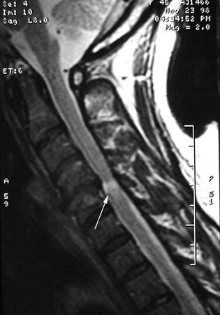
如果有神经系统体征提示超过单一神经根支配区的神经功能丧失,那么颈部 MRI 对鉴别任何神经受压的特性显得非常重要。颈部 MRI 扫描 T2 成像能够清晰地显示脊髓受压变形的情况。[3]Braga-Baiak A, Shah A, Pietrobon R, et al. Intra- and inter-observer reliability of MRI examination of intervertebral disc abnormalities in patients with cervical myelopathy. Eur J Radiol. 2008 Jan;65(1):91-8.http://www.ncbi.nlm.nih.gov/pubmed/17532165?tool=bestpractice.com [Figure caption and citation for the preceding image starts]: 在存在有症状退行性脊髓型颈椎病时,颈椎矢状位 T2 序列显示单一节段的脊髓压迫伴 T2 改变Dennis A. Turner, MA, MD [Citation ends].
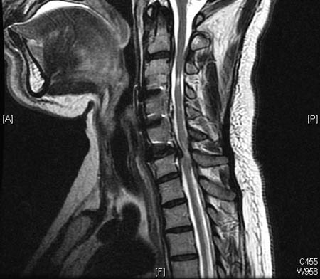
 [Figure caption and citation for the preceding image starts]: 矢状位 T2 MRI 显示前期 C3/4 水平脊髓受压,伴有残留 T2 改变;在 C2/3 和 C6/7 出现新的脊髓压迫,T2 改变Dennis A. Turner, MA, MD [Citation ends].
可以考虑使用增强或非增强颈部 MRI,以显示其他病因,包括恶性肿瘤、感染、炎性疾病及内在脊髓病或者脊髓内部损伤,即多发性硬化、肌萎缩侧索硬化。[5]American College of Radiology. ACR appropriateness criteria: cervical neck pain or cervical radiculopathy. 2018 [internet publication].https://acsearch.acr.org/docs/69426/Narrative/ 如果肌力和/或感觉丧失是单侧的,应考虑由自发性病毒性神经炎引起的臂丛神经疾病(神经痛性肌萎缩症),通常伴有颈部疼痛。
由于神经功能异常来自脊髓受压及上运动神经元功能异常,而非神经受压,对脊髓型颈椎病进行的 EMG 或 NCV 诊断性检查通常无异常,因为神经检查主要显示下运动神经元病变。然而,EMG 或 NCV 检查有助于将脊髓型颈椎病与尺神经病变或腕管综合征等周围神经病变相鉴别,这些病变可表现为许多类似症状。
无症状,但在其他疾病的诊断性评估时可能发现颈椎病。
这是一种常见表现形式。扫描结果本身被解读为异常,而且患者会试图了解扫描结果的意义。[1]Matsumoto M, Fujimura Y, Suzuki N, et al. MRI of cervical intervertebral discs in asymptomatic subjects. J Bone Joint Surg Br. 1998 Jan;80(1):19-24.https://online.boneandjoint.org.uk/doi/pdf/10.1302/0301-620X.80B1.0800019http://www.ncbi.nlm.nih.gov/pubmed/9460946?tool=bestpractice.com[4]Siivola SM, Levoska S, Tervonen O, et al. MRI changes of cervical spine in asymptomatic and symptomatic young adults. Eur Spine J. 2002 Aug;11(4):358-63.http://www.ncbi.nlm.nih.gov/pubmed/12193998?tool=bestpractice.com 患者的主诉症状通常在咨询扫描结果前缓解。
患者病史与神经功能及体格检查的关系对决定是否行进一步诊断性检查非常重要。[2]Guzman J, Haldeman S, Carroll LJ, et al. Clinical practice implications of the Bone and Joint Decade 2000-2010 Task Force on Neck Pain and Its Associated Disorders: from concepts and findings to recommendations. J Manipulative Physiol Ther. 2009 Feb;32(2 suppl):S227-43.http://www.ncbi.nlm.nih.gov/pubmed/19251069?tool=bestpractice.com[6]Binder AI. Neck pain. BMJ Clin Evid. 2008 [internet publication].https://www.ncbi.nlm.nih.gov/pmc/articles/PMC2907992/http://www.ncbi.nlm.nih.gov/pubmed/19445809?tool=bestpractice.com 如果发现有明显的神经功能异常,有可能的话应行 MRI 检查。[31]Davies BM, Mowforth OD, Smith EK, et al. Degenerative cervical myelopathy. BMJ. 2018 Feb 22;360:k186.https://www.bmj.com/content/360/bmj.k186.longhttp://www.ncbi.nlm.nih.gov/pubmed/29472200?tool=bestpractice.com 如果患者已行 MRI 检查,在决定是否对无症状患者进行治疗,尤其是当治疗可能存在特定风险时,与患者进行充分沟通很重要。
通常无症状颈椎病 MRI 结果可能包括椎体血管瘤(为先天性病变,但随时间扩大的情况罕见)、椎间盘或者面关节的退行性变(病变严重性和范围与年龄相关)、椎间盘突出、纤维环撕裂及存在可见的中央椎管(通常误认为是脊髓空洞症)。[1]Matsumoto M, Fujimura Y, Suzuki N, et al. MRI of cervical intervertebral discs in asymptomatic subjects. J Bone Joint Surg Br. 1998 Jan;80(1):19-24.https://online.boneandjoint.org.uk/doi/pdf/10.1302/0301-620X.80B1.0800019http://www.ncbi.nlm.nih.gov/pubmed/9460946?tool=bestpractice.com[4]Siivola SM, Levoska S, Tervonen O, et al. MRI changes of cervical spine in asymptomatic and symptomatic young adults. Eur Spine J. 2002 Aug;11(4):358-63.http://www.ncbi.nlm.nih.gov/pubmed/12193998?tool=bestpractice.com
 [Figure caption and citation for the preceding image starts]: 显示平均皮节面积和定位。神经根性疼痛通常局限于某一皮节。摘自美国格氏人体解剖第 20 版,经许可后使用 [Citation ends].
 [Figure caption and citation for the preceding image starts]: 在存在有症状退行性脊髓型颈椎病时,颈椎矢状位 T2 序列显示单一节段的脊髓压迫伴 T2 改变Dennis A. Turner, MA, MD [Citation ends].
 [Figure caption and citation for the preceding image starts]: 矢状位 T2 MRI 显示前期 C3/4 水平脊髓受压,伴有残留 T2 改变;在 C2/3 和 C6/7 出现新的脊髓压迫,T2 改变Dennis A. Turner, MA, MD [Citation ends].