诊断路径
当前病史
在采集红眼病现病史时,重要的是确定在更常见病因的同时是否有严重威胁视力的诊断。[1] 通过涵盖关键性问题并记下相关阴性特征,可以缩小鉴别诊断范围并且可以决定是否需要转诊以进行进一步眼科治疗,或者是否可以在初级保健机构中给予治疗。
需考虑的关键问题包括:[17]
疾病是何时开始的?
疾病是单侧还是双侧?
异物或创伤通常是单侧的,而结膜炎可从单侧开始,随后变成双侧。
症状的发作是急性的还是逐渐的?
急性发作可能提示角膜异物或磨损或者异物创伤。
患者的视力如何?
眼部是否有疼痛?
视力下降或眼内深部疼痛提示更严重的潜在疾病诊断,例如闭角型青光眼、前葡萄膜炎或巩膜炎。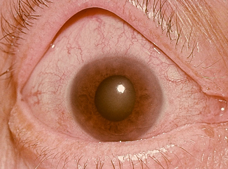 [Figure caption and citation for the preceding image starts]: 闭角型青光眼:中央角膜水肿,伴有椭圆形中度散大的瞳孔个人资料收集-由 Hugh Harris 先生提供 [Citation ends].
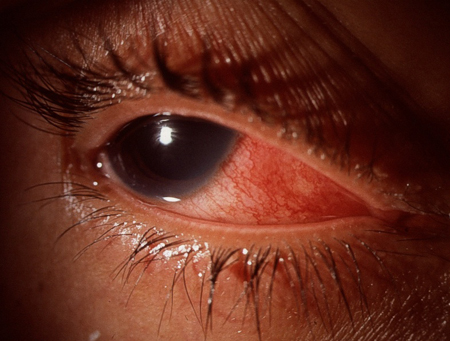 [Figure caption and citation for the preceding image starts]: 巩膜炎个人资料收集-由 Hugh Harris 先生提供 [Citation ends].
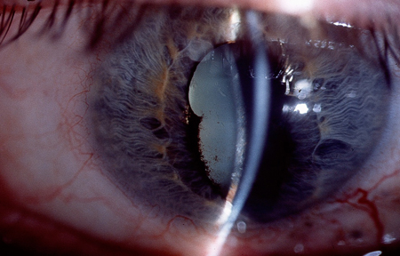 [Figure caption and citation for the preceding image starts]: 前葡萄膜炎伴后粘连个人资料收集-由 Hugh Harris 先生提供 [Citation ends].
患者存在异物感时,可能的诊断包括结膜炎、结膜/睑板下异物、角膜异物、角膜炎和角膜溃疡。怀疑有异物时,询问患者最近是否进行过可能导致此情况的任何活动。如果是,则询问是否佩戴了护目用具。活动的性质也会提示可能的穿透性损伤:例如,使用机械锯和锤可产生高速异物,这类异物能够穿透眼球表面并进入眼内。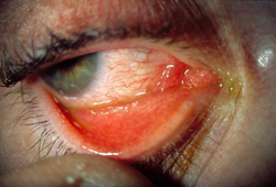 [Figure caption and citation for the preceding image starts]: 病毒性结膜炎个人资料收集-由 Hugh Harris 先生提供 [Citation ends].
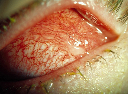 [Figure caption and citation for the preceding image starts]: 细菌性结膜炎个人资料收集-由 Hugh Harris 先生提供 [Citation ends].
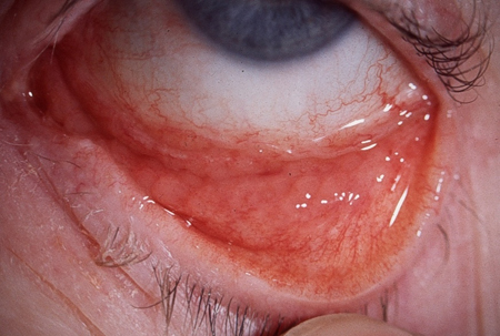 [Figure caption and citation for the preceding image starts]: 衣原体性结膜炎个人资料收集-由 Hugh Harris 先生提供 [Citation ends].
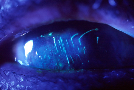 [Figure caption and citation for the preceding image starts]: 睑板下异物:通过荧光素染色可见垂直的角膜擦伤个人资料收集-由 Hugh Harris 先生提供 [Citation ends].
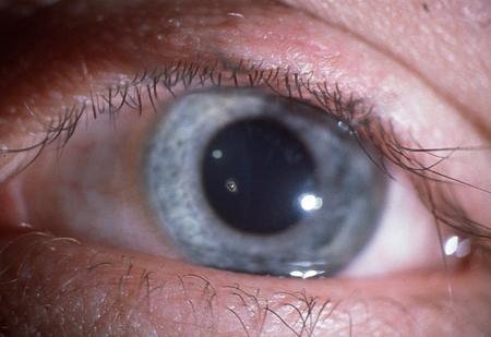 [Figure caption and citation for the preceding image starts]: 角膜异物个人资料收集-由 Hugh Harris 先生提供 [Citation ends].
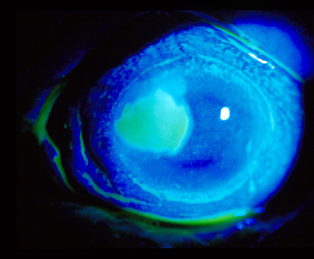 [Figure caption and citation for the preceding image starts]: 通过荧光素染色可见的角膜溃疡个人资料收集-由 Hugh Harris 先生提供 [Citation ends].
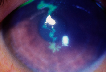 [Figure caption and citation for the preceding image starts]: 通过荧光素染色可见的树突状溃疡个人资料收集-由 Hugh Harris 先生提供 [Citation ends].
对于眼睛疼痛发红的角膜接触镜使用者,应在当天转诊进行眼部紧急评估,以避免漏诊威胁视力的并发症,如微生物感染性角膜炎或角膜溃疡。[18]
如果有任何分泌物,则能帮助识别结膜炎存在和其潜在可能病因的因素有:[19][20]
水样、脓性或黏液脓性分泌物;例如:
患病毒性结膜炎时可见水样分泌物
患衣原体性结膜炎时可见大量黏液分泌物
患淋球菌性结膜炎时可见脓性分泌物
黏液脓性分泌物可能提示细菌感染。
分泌物在早晨更严重:
可能是因为过敏反应
存在瘙痒:
通常是因为过敏反应
患衣原体性结膜炎时可出现轻微瘙痒
特应性反应的病史。
如果患者存在畏光,提示可能有潜在的前葡萄膜炎或角膜上皮损伤。对于不适的患者,务必考虑与畏光相关的全身性症状,例如脑膜炎。[21]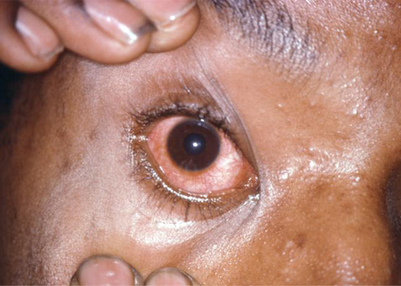 [Figure caption and citation for the preceding image starts]: 淋球菌性结膜炎CDC 图像库/Joe Miller [Citation ends].
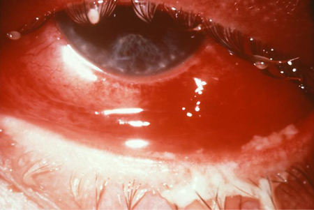 [Figure caption and citation for the preceding image starts]: 淋球菌性结膜炎:导致部分失明CDC 图像库 [Citation ends].
既往病史和既往眼科病史
医生应考虑患者既往是否出现过类似的发作,或者是否存在任何潜在的、已知可引起红眼症的相关全身性疾病,例如:
人类白细胞抗原-B27 组织相容性复合物阳性患者
反应性关节炎
结核病、梅毒
莱姆病
结节病[22]
白塞病
少关节性幼年型慢性关节炎
结缔组织病(包括类风湿性关节炎、干燥综合征和系统性红斑狼疮)
肉芽肿性多血管炎(以前被称为韦格纳肉芽肿)
复发性多软骨炎
高血压。
 [Figure caption and citation for the preceding image starts]: 结膜炎:反应性关节炎的后果CDC 图像库/Joe Miller [Citation ends].
用药史
应注意目前任何眼科药物治疗和已知会促成红眼病因形成的任何全身性药物的使用。这些药物包括散瞳剂和系统性抗胆碱能药物。服用抗凝血剂的患者可能易发生结膜下出血。局部使用抗生素但结膜炎仍持续存在时,应考虑评估其他病因。
查体
在初级保健机构,眼睛检查需要使用 Snellen 视力表、光源、荧光素和用来翻开上眼睑的棉花棒。[19] 可以使用一种分步方法,同时依据病史,考虑不同的鉴别诊断。
所有患者均应检查视力,因为视力降低可能提示红眼的一种更严重的潜在病因。
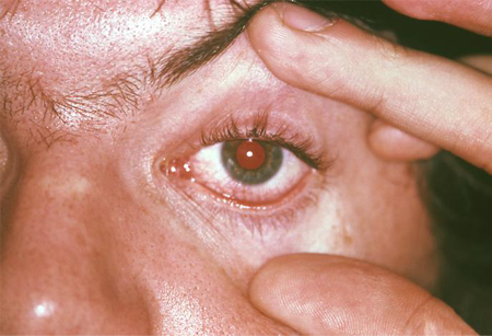
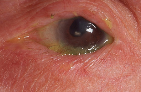
应对眼睑和眉毛进行检查,以排除眶周损伤。应检查睑缘的位置,以确定有无倒睫、眼睑内翻或眼睑外翻。如果可见任何分泌物,应考虑结膜炎的可能性。如果疾病是双侧的,并且有化脓性分泌物,那么应当作结膜炎进行治疗。眼周的水疱样皮疹可提示水痘带状疱疹或单纯疱疹感染。
 [Figure caption and citation for the preceding image starts]: 倒睫个人资料收集-由 Hugh Harris 先生提供 [Citation ends].
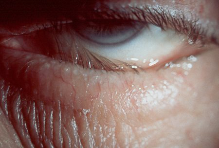 [Figure caption and citation for the preceding image starts]: 眼睑内翻个人资料收集-由 Hugh Harris 先生提供 [Citation ends].
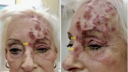 [Figure caption and citation for the preceding image starts]: 左眼带状疱疹累及前额和鼻左侧的患者(Hutchinson 征阳性;黄色箭头)。结痂的皮疹沿 V1 皮区分布,不穿过垂直中线图片经许可后使用,来自 BMJ 2019;364:k5234 [Citation ends].
在检查眼表和睑板下表面时,应对发红(一个重要特征)部位进行评估。节段性充血可能提示表层巩膜炎或存在异物。睫状或角膜缘(角膜和巩膜的交界处)充血见于前葡萄膜炎和角膜疾病。与周围结膜界限清楚的局部发红见于结膜下出血,提示应检查患者的血压。广泛性充血、较深巩膜血管充血和眼球触诊时疼痛提示存在巩膜炎。[23] 应检查睑结膜有无乳头状突起(见于过敏性结膜炎)或小囊泡(见于衣原体性结膜炎)。如果存在异物病史,那么应使用棉棒翻开上眼睑,以排除睑板下异物。如果未能发现异物并且在事故期间的活动中可能产生高速异物,那么应进一步寻求眼科意见,以排除球内的异物。在眼表检查期间滴注荧光素能够显示异物、角膜擦伤和角膜溃疡。如果角膜上出现荧光素染色或者角膜看起来浑浊(见于闭角型青光眼),那么建议转诊,以进行进一步眼科检查。当怀疑干眼为潜在病因时,可使用虎红染色。
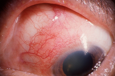 [Figure caption and citation for the preceding image starts]: 表层巩膜炎个人资料收集-由 Hugh Harris 先生提供 [Citation ends].
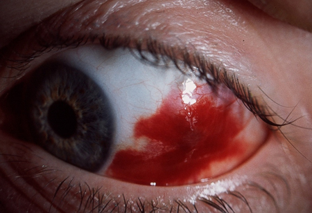 [Figure caption and citation for the preceding image starts]: 结膜下出血个人资料收集-由 Hugh Harris 先生提供 [Citation ends].
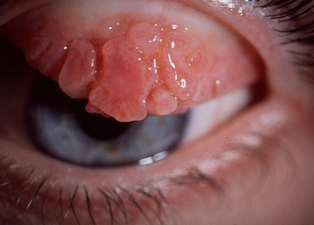 [Figure caption and citation for the preceding image starts]: 过敏性(春季)角膜结膜炎个人资料收集-由 Hugh Harris 先生提供 [Citation ends].
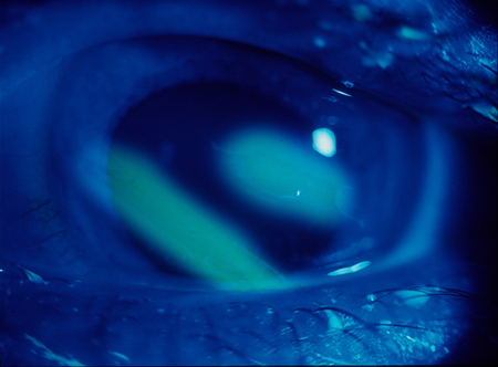 [Figure caption and citation for the preceding image starts]: 通过荧光素染色可见的角膜擦伤个人资料收集-由 Hugh Harris 先生提供 [Citation ends].
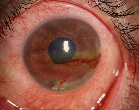 [Figure caption and citation for the preceding image starts]: 干眼(虎红染色)个人资料收集-由 Hugh Harris 先生提供 [Citation ends].
瞳孔反应。医生应观察瞳孔不等(瞳孔大小不等),如果存在这种情况,那么应转诊以进行进一步眼科评估。[20] 应使用笔形手电(或等效光源),检查直接和间接瞳孔反应。如果存在红眼,并且瞳孔反应异常,那么需排除前葡萄膜炎和闭角型青光眼。如果患者在检查时有畏光,那么也建议进一步转诊。[20]
检查
对于疑似结膜炎的患者,可以用拭子采集样本,进行细菌、病毒和衣原体培养。在给出明确的眼科诊断之后,应在专科门诊对红眼的潜在全身性病因进行检查。红眼的某些局部病因(包括睑外翻、睑内翻、角膜溃疡、角膜接触镜相关性红眼、角膜擦伤、角膜异物、巩膜炎以及闭角型青光眼)应由眼科医生进行进一步评估。穿透伤和化学伤也应由眼科医师评估。[24]
如果怀疑有高速穿透性损伤,应进行眼眶计算机体层成像影像学检查。
如果怀疑有急性青光眼,应在急诊科测量眼压。
内容使用需遵循免责声明。
